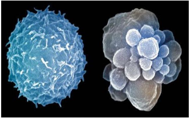
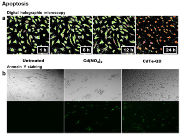
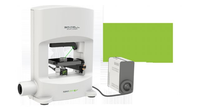
Quantum dots (QDs) are semiconductor nanocrystals with superior fluorescence properties. They have excellent performance in fluorescence labeling of cells and tissues, and can also be applied to the characterization of cell death. Recently, French scientists published a literature on the mechanism of cell death in cadmium telluride quantum dots (CdTe-QDs) in the Journal of Scientific Reports using the Nanolive label-free live cell imaging system. The researchers used rheumatoid arthritis fibroblast-like synoviocytes as subjects. First, synovial cells were treated with CdTe-QDs, and cell death was characterized by digital holographic microscopy, Annexin V staining, MDC staining, propidium iodide staining, and the like. The results show that CdTe-QDs have a toxic effect on synoviocytes and can induce cell death.     Next, the researchers used fluorescence microscopy and confocal microscopy to observe the fluorescence imaging of CdTe-QDs and to characterize and quantify the characteristics of cell death. The results showed that the fluorescence signal of CdTe-QDs in the synovial cells was significantly increased compared with the negative control. At the same time, the researchers used the Nanolive label-free live cell imaging system to image and quantify the synovial cell tomography of CdTe-QDs. The results showed that the synovial cells treated by CdTe-QDs reduced the nucleus volume by nearly half. The inner apoptotic body appears. Indicates that the cell is in the process of dying. The above results indicate that CdTe-QDs imaging can be used for the characterization of cell death.      In summary, using non-invasive Nanolive hologram label-free detection technique to determine the CdTe-QDs can be used to induce and direct visualization of the characteristics of the study of cell death, to avoid the toxic effects of traditional labeling cells, this approach has broad Application prospects, including the application of animal models, monitoring of cell-related kinetic changes, and studying the death process of other types of cells by studying the steady-state imbalance of essential metals. ★About Nanolive Labelless Live Cell Imaging (3D Cell Explorer) System★ The 3D Cell Explorer Series is a untagged, non-invasive 3D imaging system that does not conflict with any existing technology. It can reveal the most real phenomena and the most essential changes of cells, help researchers to explore from a new perspective, and help new discoveries in the field of cell life activities. It can be combined with traditional experimental methods for in-depth research, including confocal, Electron microscopy, atomic force, nuclear magnetic, mass spectrometry, etc. Since the introduction of the system in 2017, Purimai has been actively promoting the market in northern China. Currently, 3D Cell Explorer has a certain share in the Chinese market. I believe that the future market prospects must be very broad. Features: * Non-invasive or without staining marks; * Real-time monitoring of cell characteristics for up to 24 hours to several weeks; *7 channels of arbitrary digital coloring; *Nano-scale resolution; * 2 seconds of fast 3D holographic imaging; *Unlabeled IHC, IF histology and cytology studies; * Integrate 3-channel fluorescence with simultaneous 10-channel cellular information analysis (3 fluorescence + 7 digital staining = 10); Product no damage research application: * Observation and analysis of microbial infecting cells; * Mitochondrial real-time monitoring * drug mechanism and cell cycle analysis; * Yeast cell division research; *3D cell culture; * Cell autophagy research; *Nanomaterial development research; * Subcellular organelle observation; *GFP or RFP transfection analysis; * Cell interaction studies; *Botanical research; Tetanus Toxoid Vaccine,Toxoid Vaccine,Hep B Immune Globulin,Immunoglobulin Injections FOSHAN PHARMA CO., LTD. , https://www.full-pharma.com The study of cell death mechanism has been a research hotspot in the field of life sciences. In general, detection of cell death (apoptosis, autophagy, necrosis) requires indirect fluorescent labeling in conjunction with different detection methods. However, these methods do not allow real-time monitoring of internal conditions during cell death, nor can they simultaneously identify toxic substances and cell death processes. Therefore, indirect labeling is increasingly difficult to meet the needs of real-time monitoring of cell death processes.


