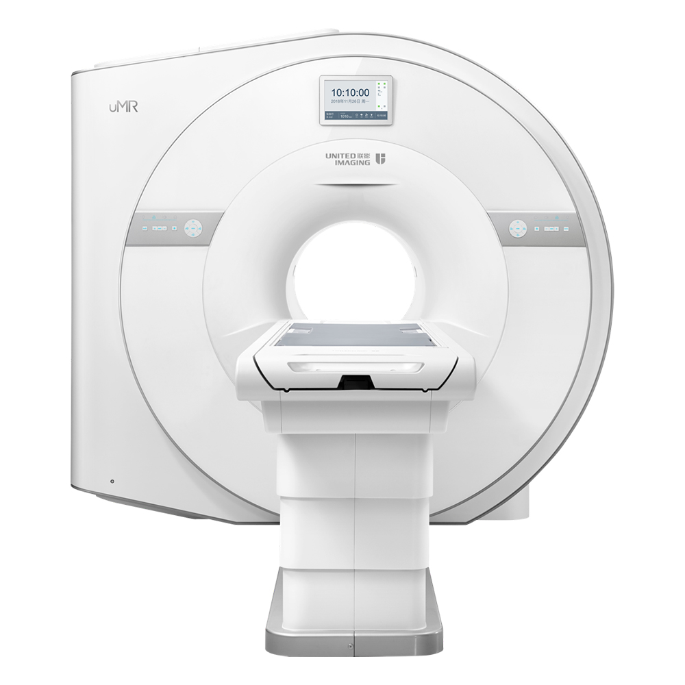
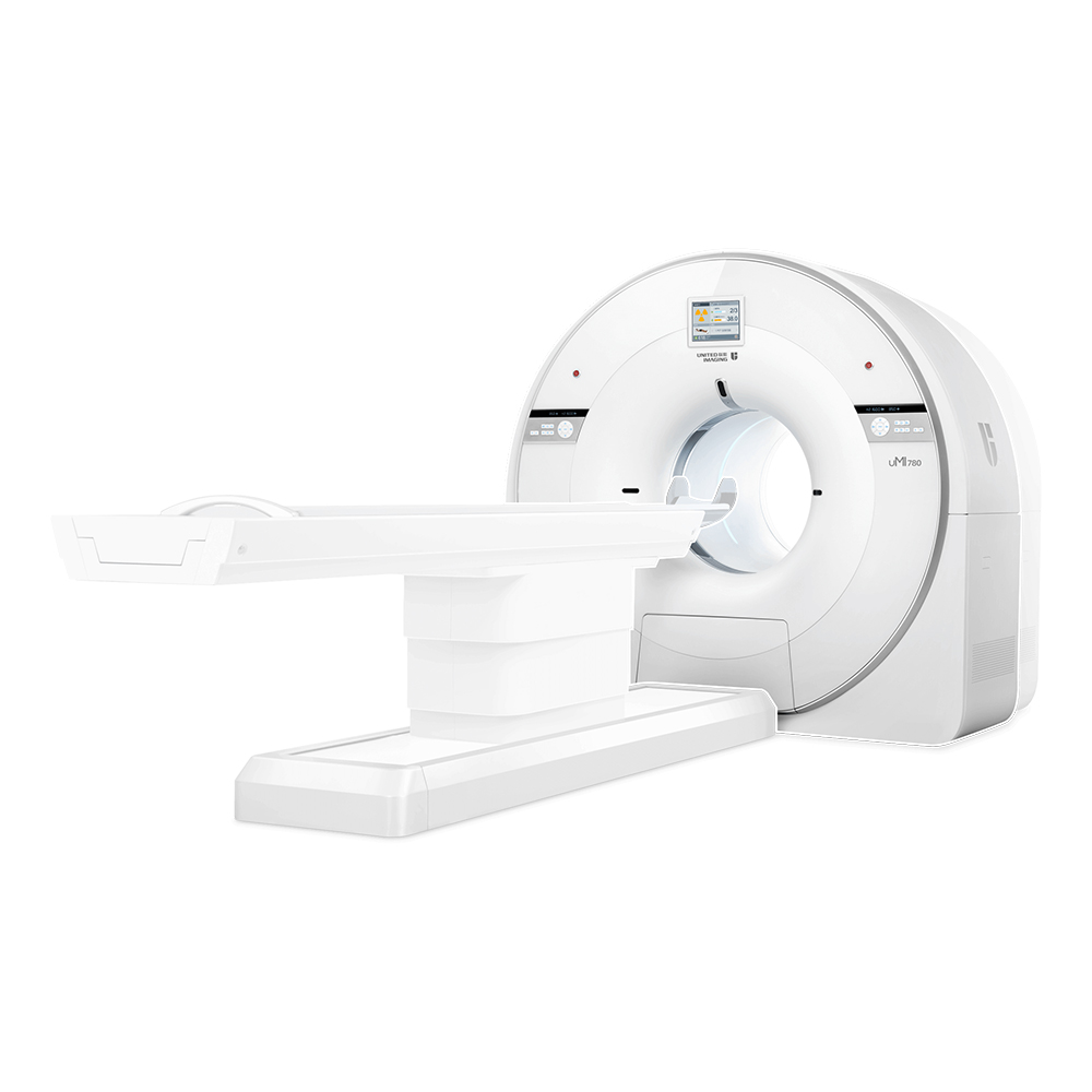
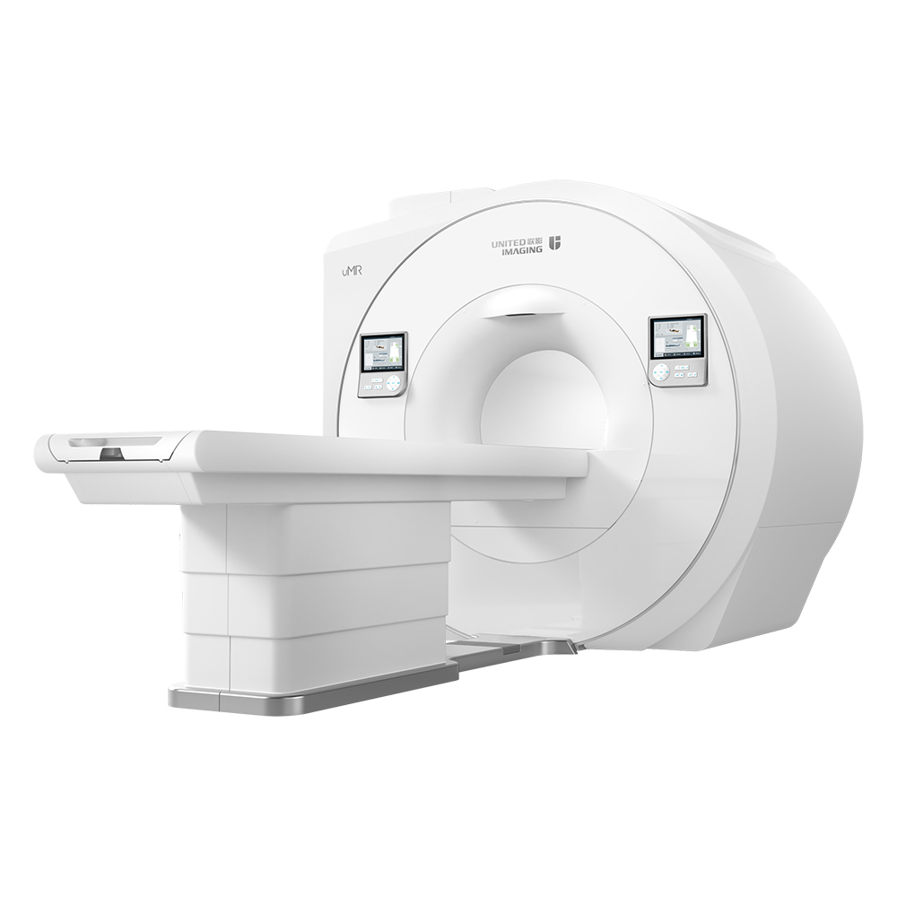
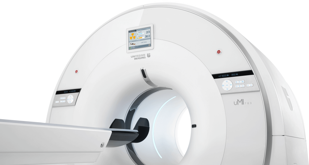
A CT scan makes use of computer-processed combinations of many X-ray images taken from different angles to produce cross-sectional (tomographic) images (virtual "slices") of specific areas of a scanned object, allowing the user to see inside the object without cutting.
Digital Radiography made affordable, Created to meet the needs of community to hospitals and private radiology practices, it enables the price-sensitive customers to join the drive to go digital.The versatile floor mounted radiography system enables you to go from film to CR to DR.Flooe-mounted easy for installation and operation
Computed Tomography,Medical Computed Tomography Scanning Machine,Medical Ct Scanner,Computed Tomography In Ct Scanner Shanghai Rocatti Biotechnology Co.,Ltd , https://www.ljdmedical.com
This DNA "molecular cage" was developed by Oxford University physicists and molecular neuroscientists. It consists of four synthetic DNA short chains that can self-assemble into a tetrahedron about 7 nanometers tall (4 by The triangular pyramid forms).
In previous studies, Oxford researchers have demonstrated that these short chains can be assembled around protein molecules to form a "molecular cage" that encloses the protein and can be designed to allow "molecular cages" to encounter The specific "trigger" molecule in the cell opens again.
In the new experiment, they introduced fluorescently labeled DNA tetrahedron "molecular cages" into laboratory cultured human embryonic kidney cells and found that most of these "molecular cages" were intact and at least resistant to cellular enzyme attack for 48 hours. as long as. This viability is critical for drug delivery tools. DNA "molecular cages" must be able to efficiently enter cells and survive until they are released at the right time and place.
Andrew Tupperfield, a professor of physics at the University of Oxford who led the study, said: “We have tested the ability to manufacture and control DNA cages. This is important, but it is only the first step. More importantly, these cages can carry The goods or drugs enter the interior of living cells."
Teporfield explained that according to previous studies, the size of delivery tools is an important factor in determining whether they can enter cells successfully. If the diameter is less than 50 nanometers, it is more likely to enter the interior of cells than larger particles. This tetrahedron "molecular cage" only 7 nanometers, not only is easy to enter the cell, but also left a considerable space to accommodate useful goods. There is still a need to further study the path of these DNA "molecular cages" into living cells.
The United-imaging is floor mounted system comprises a radiographic table with integral floor guide rail and a wallstabd. Requiring little room preparation, it is easy to install.
United-imaging according to the international advanced processing mode and standardlize design. the parts are made with precision CNC machine and moulding. Which adopt high degree of standardized, reasonable and compact structure. With reliable, durable and elegant appearance and advanced processing technology.


DNA "molecular cage" can be made into nano-scale drug delivery vehicles
Recently, Oxford University scientists have for the first time developed a molecular "cage" made from DNA that can enter and survive inside living cells, which may lead to an effective new drug delivery method. Research papers were published in the electronic journal ACS Nano.