Whole tissue cytochrome oxidase activity staining kit product manual (Chinese version) The main purpose Whole tissue cytochrome oxidase activity staining reagent is a non-enzymatic natural oxidation of cytochrome C catalyzed artificial electron donor dye diaminobenzidine to form a brown insoluble product to analyze cytochromes in whole tissue samples. An authoritative and classic technical approach to oxidase activity. This technology has been proven through careful improvement of traditional NADI methods and successful experiments. It is mainly suitable for the qualitative detection of cytochrome oxidase activity in various animal tissues. The product is strictly sterile, ready to use, simple in operation, stable in performance and clear in color. technical background Cytochrome oxidase is a general term for the enzyme portion of the oxidative respiratory chain. It is present on the mitochondria of eukaryotic cells and supplies energy to cells primarily through oxidative phosphorylation. Diaminobenzidine (DAB) is an artificial electron donor dye that produces non-enzymatic natural oxidation while providing electrons by physical binding to metalloproteins such as ferritin-containing cytochrome C. (Cytochrome C acts as an electron acceptor) in the electron transport system of the respiratory chain, thereby exhibiting color changes and deposition, forming a tan insoluble product, and is not affected by ethanol. At the same time as the DAB is naturally oxidized, active oxygen, such as hydrogen peroxide, is generated, which is prevented from accumulating by catalase. In the presence of a large amount of oxidized DAB, ferrous cytochrome C is oxidized to normal iron cytochrome C by the action of cytochrome oxidase. The oxidized DAB tan insoluble product was thus used to detect the activity of cytochrome oxidase in tissue cells. product content Cleaning solution (Reagent A) 200 ml Dyeing Liquid A (Reagent B1) 40 ml Dye B (Reagent B2) 4 bottles Reaction solution (Reagent C) 3 ml Fixative (Reagent D) 30 ml Product manual 1 copy storage method Store the staining solution (Reagent B) and the reaction solution (Reagent C) in a -20 °C refrigerator to avoid light; the rest are stored in a 4 °C refrigerator to ensure June. User-supplied 1.5 ml centrifuge tube: container for working fluid preparation Incubator: for reaction incubation Neutral resin: used for slicing Optical microscope: used for observation and analysis after sectioning Experimental procedure Before the start of dyeing, melt the reagent in the -20 °C refrigerator at room temperature; remove 9 ml of staining solution A (Reagent B1) into 1 bottle of staining solution B (Reagent B2) , mix and mark as dyeing working solution . Place in the ice tank for use, avoiding light (note: valid for 1 week); continue to transfer 2.7 ml of dyeing working solution to a new 1.5 ml centrifuge tube, add 300 μl of reaction solution (Reagent C) , mix and mark, mark The reaction working solution is placed in an ice bath to avoid light. Then do the following. Precautions The company provides a series of tissue cell enzyme activity dyeing reagent products Quality Standard
pharmaceutical blister Packaging
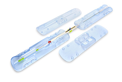
with excellent transparency and smoothness, display effect is good. the surface decoration performance is excellent, can be printed without surface treatment, easy to press the pattern, easy to metal treatment (vacuum gold-plated layer) . has good mechanical strength. Good barrier performance for oxygen and water vapor. good chemical resistance, can withstand a variety of chemical substances corrosion. non-toxic, reliable health performance, can be used for food, medicine and medical equipment packaging, and can be Y ray [SPAN] to the packaging of the goods. good adaptability to environmental protection, can be economical and convenient recycling; When the waste is incinerated, it does not produce harmful substances harmful to the environment.
Pharmaceutical Packaging,Pharma Blister Packaging,Blister Packaging Medication,Pharmaceutical Blister Packaging taicang hexiang packaging material co.,ltd , https://www.medpackhexiang.com