Release date: 2016-09-19 X-ray transmission, commonly known as "shooting X-ray film", is a medical diagnostic technology that everyone is familiar with. X-ray energy is high and can penetrate the skin, so taking X-ray film is the standard practice for doctors to diagnose bone injury. Hospital X-ray doses are generally safe, but even so, if the patient's short-term class requires frequent bone imaging, this high-energy radiation can have adverse effects on the body. Source: X-MOL Natural Sweetener Polyglucan,Sugar Replacement Polydextrose,Better Fiber Polydextrose Shandong Bailong Chuangyuan Bio-tech Co.,Ltd. Qingdao Branch , https://www.sdblcycn.com
Professor Thorfinnur Gunnlaugsson from Trinity College, University of Dublin, in collaboration with scientists from the Royal College of Surgeons of Ireland, invented a revolutionary high-resolution 3D skeletal imaging technique to replace X-ray transmission. The team's work was published in Cell Press's newly published journal Chem. (Two-Photon Luminescent Bone Imaging Using Europium Nanoagents. Chem, 2016, 1, 438-455, DOI: 10.1016/j.chempr.2016.08.011)
 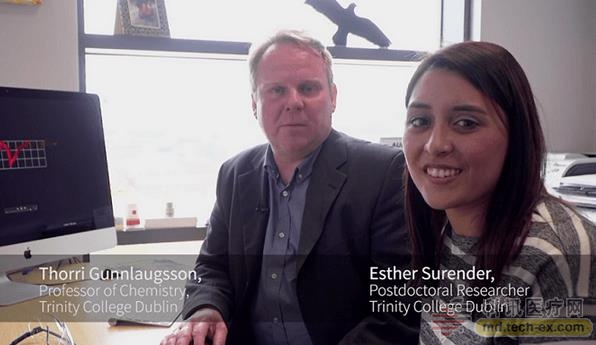
Professor Gunnlaugsson (left) and this article are Dr. Esther M. Surender. Image source: Trinity College Dublin
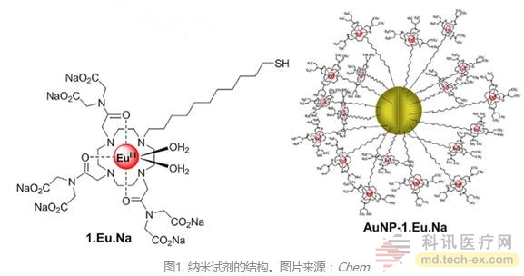
The scientists coordinated complex polydentate ligands with trivalent europium ions and combined the complexes with gold nanoparticles with a thiol-containing linker at the end to form a nanocompatible reagent with very good biocompatibility. They are automatically enriched on calcium-rich surfaces – often tiny gap wounds on the bones. These nanoparticles are therefore able to target and mark cracks on the bone, helping researchers to create a complete three-dimensional map of the injured area's bones.
This technology has a huge impact on medicine, it can diagnose the strength of bones and provide all the information about bone injuries. In addition, the technology can also serve bone graft surgery and can be used as an early warning system for degenerative orthopedic diseases such as osteoporosis.
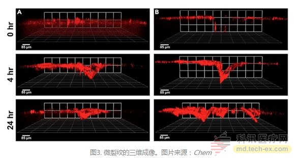
Professor Gunnlaugsson said: "This outstanding work has benefited from the collaboration of chemists at Trinity College with RCSI medical experts and engineering experts. We have demonstrated that this new three-dimensional fluorescence imaging technology can display so-called microcracks. Nanoparticles provide unprecedented images and information. This article is an important step in our clinical application to the diagnosis of bone injuries."
 
Professor T. Clive Lee, one of the authors of the article, believes that “our daily activities will put a burden on the bones and lead to an increase in microcracks. Under normal circumstances, the bones will repair these cracks themselves, but if microcracks It develops too fast, and beyond the speed of repair, it can make bones fragile. This often occurs in athletes and causes stress fractures. For the elderly with osteoporosis, bone repair is slow, microcrack development Rapid results in fragility fractures, especially in the buttocks, wrists, and spine. Current X-ray techniques provide bone morphology but do not give information about bone quality."
He continued. "We used new nanoparticles to mark microcracks and probe them with magnetic resonance imaging (MRI). We also examined bone mass and bone mass, located where the fracture was most likely to occur, and developed a treatment plan. Diagnosing potential problems before fractures can reduce orthopedic surgery and transplantation – prevention is better than cure.â€
In addition to powerful imaging capabilities, another obvious advantage of this technique is that the patient does not need to be exposed to X-rays. A dose of X-ray radiation can increase the risk of cancer in some cases. The red gold nanoparticles used in this technology are very biosafe. In fact, gold has been safely used in a variety of medical treatments.
Dr. Surender believes that these nanoparticles have good clinical application prospects. First, gold nanoparticles can reduce the concentration of reagents that need to be injected into the body, which is beneficial for clinical use. Second, the long-wave radiation used to stimulate imaging is essentially harmless to the organism.
These nanoparticles are generally similar to the currently used MRI contrast agents, but if strontium ions are used instead of strontium ions, MRI can be combined with X-ray and computed tomography (CT).
Coincidentally, the Trinity College of Biomedical Research, where Professor Gunnlaugsson is located, celebrates its fifth anniversary. He lost no time in “selling†his research to top experts in chemistry, immunology, bioengineering and cancer biology from around the world.
Â
1. http://
2. https://