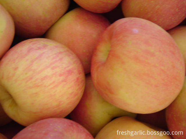
Fuji apples are typically round . Fresh apples contain between 9–11% sugars by weight and have a dense flesh that is sweeter and crisper than many other apple cultivars, making Fuji apples popular with consumers around the world. Fuji apples also have a very long shelf life compared to other apples, even without refrigeration. With refrigeration, Fuji apples can remain fresh for up to a year.
1. Commodity name: Fuji Apple
Fuji Apple,Fresh Apple,Red Fuji Apple,Fresh Fuji Apple JINING FORICH FRUITS & VEGETABLES CO., LTD. , https://www.forichgarlic.com
3. Coloration: 80%-85% and up, color type seperated with blush or strip, smooth and bright skin
4.Origin: Shandong province of China
5. Packing:
a) Inner packing: With tray, foam net and plastic bag
b) Outer packing:
10kg/ctn: size 28/32/36/40/44/50/56;
20kg/ctn: size 64/72/80/88/100/113/125/138/150/163/175/198;
c) according to clients' special requirements.
6. Supply Period: October to next August
7. Conveyance:
a) 10kgs/ctn: 2156ctns/40' HR
b) 20kgs/ctn: 1106ctns/40' HR
8. Transporting and storing temperature: 0°C

Long-term behavioral testing of neonatal rats after hypoxic-ischemic brain damage
Long-term behavioral testing of neonatal rats after hypoxic-ischemic brain damage
[Abstract] Objective At present, the pathological and biochemical indicators of the hypoxic-ischemic brain damage (HIBD) model in neonatal rats are often used, but lack of functional evaluation methods. This study explored the behavioral changes and methods of determination of HIBD in neonatal rats, and provided a functional evaluation method for the study of HIBD in neonatal rats.
Methods Twenty-four newborn Sprague-Dawley rats aged 7 days were randomly divided into control group (n=12) and HIBD group (n=12). Rats in HIBD group received hypoxia-ischemia, and rats in control group only received sham operation. Three weeks after birth, the two groups of rats were evaluated by T-maze test and sensorimotor test for learning, memory, spatial ability and sensorimotor function. Then, the two groups of rats were sacrificed, and the brain tissue sections were taken for Nissl staining. The number of neurons per unit area of ​​hippocampal DG and cortical areas was counted, and the behavioral and histological results were analyzed.
Results In the behavioral test, the scores of the rats in the HIBD group were significantly lower than those in the normal rats. In the T-maze test, there was a significant difference between the two groups at the 3rd and 4th (P=0.049, P<0.001), and the correct rate of the HIBD group (68.3%±26.2%, 66.7%±15.6%) was significantly lower than that. Control group (86.7% ± 15.6%, 98.3% ± 5.7%). In the sensorimotor test, compared with normal mice, HI BD rats showed asymmetry of left and right motion in both foot error and limb placement tests (both P < 0.001), and posture reflexes also showed motor dysfunction (P = 0 . 032). Hypoxia-ischemia caused neuronal damage, resulting in hippocampal DG (39. 7 ± 5. 9 vs 50. 9 ± 4. 1 , P < 0.001) and cortical unit area neurons (12.7 ± 3) 3 vs 18. 2 ±3 . 3, P < 0 . 001) The number is significantly reduced. However, behavioral test results were not associated with histological changes.
Conclusion Hypoxic-ischemia in neonatal rats can cause long-term learning and memory, spatial ability and sensorimotor dysfunction. T-maze test and three sensorimotor tests can be used as evaluation indexes of rat HI BD model.
[Key words] hypoxia-ischemia, brain behavior, neonatal rats
Hypoxic ischemic encephal opa2thy (HIE) is the most common cause of brain injury in the neonatal period, and is also a common cause of cerebral palsy, mental retardation, learning disabilities and epilepsy [1].
The hypoxic2ischemic brain damage (HI BD) model of neonatal rats prepared by Rice method was made by unilateral carotid artery ligation in 7-day-old neonatal rats and exposed to hypoxia. 7-day-old rats are at the peak of brain development. At this time, brain damage caused by hypoxia-ischemia is similar to that of full-term brain injury caused by perinatal asphyxia [2], which is the study of H IE injury mechanism and protective factors. Common animal models. In addition to reducing neuronal damage, treatments that protect HIBD should, more importantly, improve long-term functional changes caused by brain damage. However, the current research on the protection of HIBD mostly uses pathological and biochemical indicators, and less evaluation of long-term function. In the central nervous system, especially in immature brains, tissue repair and compensatory mechanisms affect long-term damage and functional recovery, so recent pathology or behavioral assessments are not necessarily consistent with long-term assessments [3, 4 ].
HIBD is a diffuse brain injury that can cause unilateral cortex, striatum and hippocampal damage [2,5]. Cortical damage can lead to impaired sensorimotor function [4]; striatum injury can lead to changes in spontaneous movement [5], while hippocampal injury leads to decreased spatial memory and learning ability [6]. However, in the neonatal rat HIBD model, striatum injury only leads to recent spontaneous motor changes, which will be restored after weaning [5]. Therefore, this study evaluated the long-term learning and memory, spatial ability and sensorimotor function of rats after hypoxia-ischemia with reference to the behavioral test methods of Balduini [5] and Bona [4], providing an evaluation method for the intervention study of HIBD. .
1 Materials and methods
1.1 Animals and grouping
Fresh-cleaned Sprague 2 Dawley rats, male or female, weighing more than 12 g, were randomly divided into control group and HIBD group, with 12 rats in each group. Newborn rats were breast-fed by female rats, weaned at 21 days of age, and male and female were caged. Clean and drink water, maintain room temperature at 22 ° C, give 12h light, 12h dark.
1.2 HIBD model making
In the HI BD group, the rats were inhaled and anesthetized at 7 days of age. The left common carotid artery was isolated, ligated and disconnected. The skin was sutured and returned to the mother cage for 2 hours, and then placed in an 8% nitrogen-oxygen mixture. °C incubator for 2 h. The rats in the control group underwent sham operation at 7 days of age, and only the left common carotid artery was isolated without ligation and hypoxia.
1.3 Behavioral testing
Both are performed in a double-blind manner, that is, the tester does not know the grouping of animals.
1 . 3 . 1 T maze test T maze is wooden, each arm is 11 cm wide, 18 cm high, 40 cm long, and has a gate at 15 cm from the end of the starting arm. Tested from the age of 22 days. A 5 cm diameter vessel was placed at the end of the two arms of the T-maze, and 40 mg of food was built in. The test animals were tested after 2 days of foraging in the T-maze. The test is divided into two stages of preparation and testing. In the preparatory stage, arbitrarily select one arm of the T-maze, close it with a wooden gate, the animal can only enter the open arm, and eat the food, then put it into the starting arm, and then remove all after 15 s. The gate began testing. At this stage, the animal's hind limbs entered either of the arms and considered to have made a choice and were not allowed to retreat. If the animal enters the arm that has not entered the preparatory stage, it is allowed to return to the cage after eating the food; if it enters the arm that has already entered, it is returned to the cage after being closed for 10 s. Each animal was tested 5 times a day for 15 minutes each time for 4 days. Record the correct rate of daily testing.
1 . 3 . 2 sensory motor function test ( sens ori mot or functi ontest) includes the following three tests. The test was performed when the rats reached 34 days of age.
1 . 3 . 2 . 1 foot2 faults test Place the rats on a horizontal metal grid (50cm × 40cm, 3cm × 3cm per cell, metal diameter 0.4cm), record 2min claws fall into the grid The number of times was to exclude the influence of the difference in the activity of different rats, and only the difference between the number of errors on the left and right sides was statistically analyzed.
1 . 3 . 2 . 2 Postural reflex test Grasp the tail of the rat and hang it at a height of 50 cm from the table top. Normal rats extended both forelimbs to the table top (0 points), while rats with brain damage damaged the limbs on the opposite side (right side) of the cerebral hemisphere (1 point), and then placed the rats on the table. Pressurize the side of the shoulder until the forelimb is straightened, repeating several times, if the damage to the opposite side (right side) of the damaged hemisphere is reduced to abnormal (2 points).
1 . 3 . 2 . 3 limb placement (li mb2 p lacing test) Record the placement of the left and right hind paws of the rats under different sensory stimuli. The scoring criteria are as follows: 0 points, the paws are placed correctly and quickly; 1 point, Slow or not completely correct; 2 points, can not be placed. The difference in scores on both sides of each mouse was recorded. 1 Place the rat on the table. Normally, the rat will stretch the forepaws on the table. 2 Raise the rat's head with your fingers to avoid seeing the table, and touch the forelimbs of the rats to the edge of the table to detect the feeling of the forelimbs. 3 Rats were placed on the edge of the table to detect the placement of the forelimbs. Normal rats placed the double front paws on the table; while the brain injury rats placed the side paws incorrectly; 4 The examiner moved the rats slowly to the edge of the table to detect the placement of the front and rear limbs; 5 Place the rats on the table On the top, gently push it from the side to the edge of the table, the normal rat will grab the edge of the table, and the front and back limbs of the opposite side of the brain injury rat may fall; 6 with 5, just push from the back.
1.4 Tissue sectioning and staining
Four weeks after HIBD, the rats were sacrificed, 4% paraformaldehyde was perfused, and brain tissue was fixed and embedded in paraffin, and 3 μm paraffin sections were prepared. After dewaxing and hydration, Nissl staining was performed. Nissl staining was performed at 3 mm from the anterior and posterior iliac crests, 0.1% toluidine blue at 60 ° C for 1 min, washed with water, 70%, 80% alcohol dehydrated, and differentiated by 95% alcohol until the nucleus and background were light blue or Colorless, 100% alcohol dehydrated, turpentine transparent, sealing. Under the light microscope (×200), neurons were counted per unit area at the same location.
1.5 Statistical analysis
All data were processed using SPSS11.0 software package statistical software. The measurement data is expressed as x ± s, and the mean is compared using the t test; the grade data is compared using the Mann2 whitney U test. Correlation analysis was based on pears on correlation analysis (measurement data) or s pearman correlation analysis (grade data).
2 results
2.1 Behavioral testing
2 . 1 . 1 T labyrinth test in each group of tests for 4 consecutive days, the correct rate is shown in Table 1. With the increase of the number of days, the correct rate of the control group gradually increased, while the correct rate of the HIBD group did not increase significantly, indicating that the learning ability was poor. At the 3rd and 4th day, the difference was significant.
2 . 2 sensory motor function test
2 . 2 . 1 The difference between the right and left of the HIBD group (3.0±1.0) was significantly higher than that of the control group (0.7 ± 1.3) (t = 2 4 . 841, P <0 . 001), indicating that The left and right movements of the rats in the group were asymmetric, and the right side (opposite to the ischemic brain) was worse than the left side
2 . 2 . 2 Posture Reflexes Scores of rats in each group in the posture reflex test. The higher the score, the more serious the brain damage. In the control group, 12 cases were 0 points, while in the HI BD group, only 8 cases were 0 points. The difference between the two groups was significant (Z = 2 2 . 145, P = 0. 032).
2 . 3 Nissl stain
Hypoxic-ischemic ischemia resulted in neuronal damage, resulting in a significant reduction in the number of neurons in the hippocampal DG and cortical unit area (both P < 0.01) (Table 3, Figure 2).
2.4 Correlation analysis of histological and behavioral results The correlation between the number of neurons per unit area of ​​the hippocampal DG area of ​​HI BD rats and the correct rate of the fourth day of the T-maze test was not correlated (r = 2 0 043, P = 0 . 896 ). There was no correlation between the number of neurons per unit area and the scores of the three sensorimotor scores in the HI BD rat cortical area (rs = 0.02, 286, P = 0. 368).
Source: http://