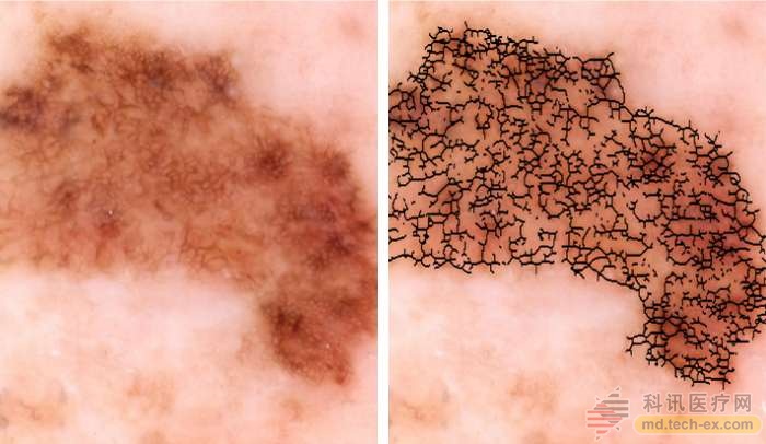
Release date: 2016-12-28 Even experts may be fooled by melanoma. People with this type of skin cancer often look like cockroaches on the skin, are often irregular in shape and color, and are difficult to distinguish from benign, making the disease difficult to diagnose. Now, Rockefeller University researchers have developed an automated technology that combines imaging with digital analysis and machine learning to help doctors detect melanoma early. “The dermatology field really needs to standardize how to evaluate melanoma,†said Professor James Carl, director of the clinical investigation and investigation dermatology laboratory at Martin Carter. “Screening tests can save lives, but it is visually challenging, even if When suspicious lesions were extracted and biopsied, only about 10% were confirmed to be melanoma." In the new method, the lesion image is processed by a series of computer programs and other quantitative data extracted about the color and quantity. This analysis yields an overall risk score, called the Q score, which indicates the likelihood of cancer. The study was published in experimental dermatology, and a recent evaluation study showed a Q-score sensitivity of 98%, which means it is likely to correctly identify early melanoma on the skin. The ability to test a correct diagnosis of normal sputum was 36%, close to the level achieved by an expert dermatologist performing a visual examination of the suspect under the microscope. The first author, Kruger Laboratories' clinical investigation report and teacher, Daniel Gareau, said: "The success of the Q score in predicting melanoma is a significant improvement in competitive technology." The researchers developed the tool by providing 60 photos of cancer melanoma and an equal amount of benign growth photos to an image processing program. They developed imaging biomarkers to accurately quantify visual features. Using the calculation method, they produced a set of quantitative metrics that differed between the two sets of images and gave each biomarker a malignant rating. By combining the data from each biomarker, they calculated the total Q score for each image, between 0 and 1, with a higher number indicating a higher probability of cancer. As shown in previous studies, the color in the lesion proved to be the most important biomarker for determining malignancy. And some biomarkers are only correct when viewed in a specific color channel - the researchers say these findings may be exploited to identify other biomarkers and further improve accuracy. “I think this technology can help detect diseases early, which can save lives and avoid unnecessary biopsy,†Gareau said. “Our next step is to evaluate this method in a larger study and take a closer look. How do we use specific color wavelengths to reveal lesions that may be invisible to the human eye but still available for diagnosis." Source: Noble Qc Test,Blood Glucose Qc,Blood Glucose Test Qc,Glycohemoglobin Qc Solution Wuxi BioHermes Bio & Medical Technology Co., Ltd. , https://www.biohermesglobal.com